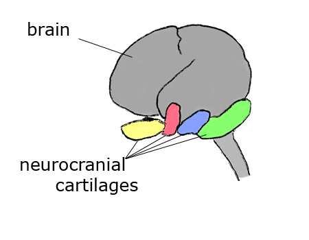
The neurocranium is a region of the head skeleton which forms from cartilages that develop around the floor of the brain just anterior to the notochord. The image below depicts the approximate locations of the neurocranial cartilages in human fetal development.
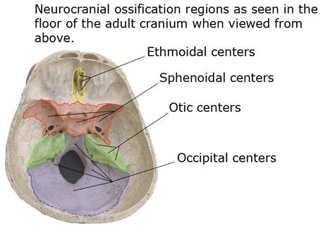
Later, the cartilages form the floor of the cranium from the ethmoid, near the anterior end, to the occipital, surrounding the foramen magnum at the posterior end.
These neurocranial bones become tightly fused with dermatocranial and splanchnocranial parts in the fully developed skull.
The neurocranial elements begin as disconnected embryonic cartilages. One pair of cartilages lies ventral and lateral to the posterior brain region. These are the parachordal cartilages. Another pair of cartilages, the prechordal cartilages, lies ventral and lateral to the brain just anterior to the parachordals.
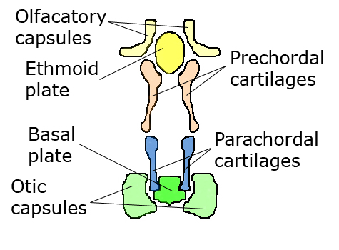
The prechordals become connected to the median ethmoid cartilage near the anterior end of the developing brain, and the parachordal cartilages become connected to the basal plate cartilage near the posterior end of the brain. These six cartilages form a supportive base at the perimeter of the developing brain. In addition to this supportive base, additional cartilages begin development around the paired internal ear structures and olfactory organs. These become the otic capsules and olfactory capsules, respectively. In most species all of these cartilages become fused or tightly connected into all or part of the brain case during skull formation. In the human these elements become the ethmoid bone, part of the sphenoid bone, part of the temporal bones, and part of the occipital bone.